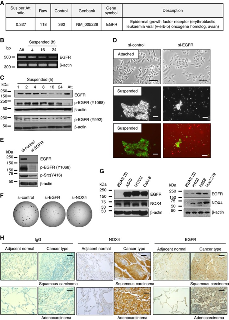
Figure 6.
Association of EGFR levels with anoikis sensitivity in A549 cells. (A) A549 cells were grown in attached or suspended conditions for 24 h, and total RNA was extracted and processed for expression microarray. (B and C) A549 cells were grown in attached or suspended conditions for 24 h or indicated times, followed by total mRNA extraction for RT-PCR (B) and cell lysate preparation for immunoblotting analysis (C). (D and E) A549 cells were transfected with si-control or si-EGFR RNAs and were grown in attached or suspended conditions for 24 h, followed by cell viability analysis (D) or immunoblotting analysis (E). (F) A549 cells were transfected with si-control, si-EGFR, or si-NOX4, and 1 × 10³ cells/well of a 35 mm culture plate were seeded and cultured in soft agar medium for 2 weeks, followed by staining with INT and imaging. (G) Cell lysates from BEAS-2B, A549, H1703, Calu-6, H460, H358 and HCC2270 cell lines were analysed by immunoblotting using anti-EGFR and anti-NOX4 antibodies. Similar results were observed in three independent experiments. (H) A patient-derived lung cancer tissue microarray was examined for NOX4 and EGFR expression using an immunoperoxidase method and isotype IgG was used as a control for staining. The staining results were graded according to intensity and proportion of positive cells described in the ‘Materials and Methods' section.

