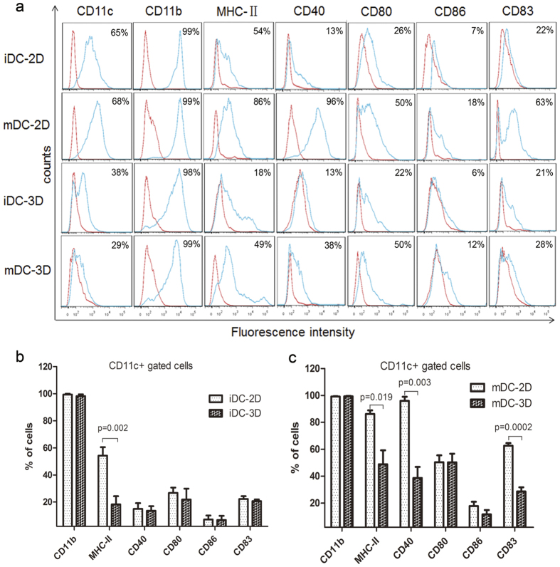
Figure 2. Immunophenotypic analyses of DCs cultured in 2D and 3D collagen scaffolds by FACS.
(a) Phenotypes of iDCs-2D, mDCs-2D, iDCs-3D, and mDCs-3D. DCs differentiated in 2D and 3D collagen scaffolds were stained using Abs specific for CD11c, CD11b, MHC-II, CD40, CD80, CD86, and CD83 as described in the Materials and Methods. Red lines represent cells stained with isotype-matched control Abs. Statistical analysis of the surface antigen molecules in iDCs-2D and iDCs-3D (b) and mDCs-2D and mDCs-3D (c). Data are representative of three independent experiments (mean ± SD). **P < 0.01 versus iDCs-2D (b). *P < 0.05, **P < 0.01, and ***P < 0.001 versus mDCs-2D (c).

