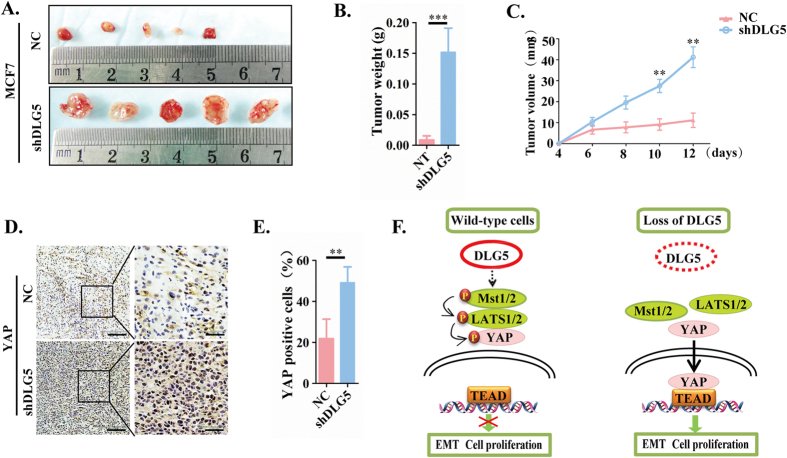
Figure 6. Loss of DLG5 induces tumor proliferation in vivo.
(A) NC or MCF 7-shDLG5 cells (5 × 10⁶) were subcutaneously injected into nude mice. Tumors were excised, and images were taken after 12 days. Tumor weight (B) was measured after 12 days, and tumor volume (C) was recorded every two days after tumor inoculation. (D). YAP and SCRIB localization was analyzed by immunostaining. Low-power scale bar, 150 μm; high-power scale bar, 50 μm. (F) Schematic of the role of DLG5 in breast cancer progression to malignancy.

