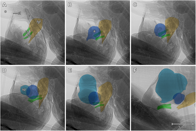
Figure 3. Radiography of male and female genital interaction during spermatophore transfer by a bushcricket mating couple.
The images were extracted from the video S2, which was recorded at recorded at 25 fps, 3.6x magnifications, 3.06 μm pixel size. (A) During the spermatophore ejection the male’s cerci (c) help to keep the female’s and male’s genitalia in close contact. (B,C) The male’s titillators (t1 and t2, green) are pushed down on the female’s genital fold (fgf, yellow) during the ejection of the ampullae (a, dark blue). (D–F) While pumping out the spermatophylax (sp, light blue), the ampullae slide over the titillators and are attached on the female’s genital opening.

