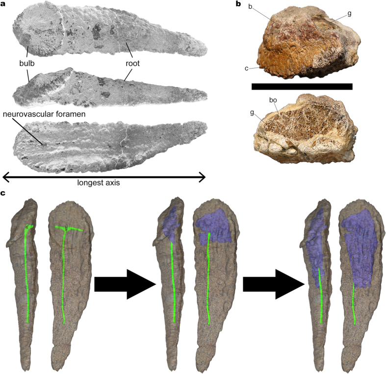
Figure 1. Titanosaur bulb and root osteoderms anatomy.
(a) Basic bulb and root osteoderm anatomy shown in superficial (top), lateral (middle), and deep (bottom) views. (b) Fragmentary osteoderm HUE-08738 in lateral view (top), and its section (bottom), showing large trabeculae with chaotic pattern filled with gypsum within most of its volume, revealing the ultrastructure of titanosaur osteoderm internal large hollow spaces (reproduced from Vidal et al. 20143). (c) 3D model of the internal canals based on the information revealed by fossils and a hypothetical transformation into large hollow spaces based on the evidence from HUE-00950 and HUE-02000. The low density bone appears in the largest volume, under the bulb region, and develops toward the root end. c = cingulum. b = bulb. bo = bone. g = gypsum.

