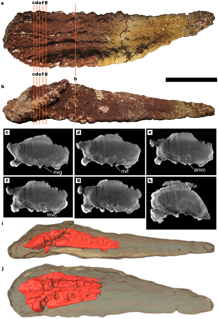
Figure 4. Titanosaur osteoderm with large internal cavities.
(a) HUE-02000 in deep view. (b) HUE-02000 in lateral view. (c–h) CT-scan slices at different positions, where the lower density area and the neuro-vascular canal can be observed. Notice how the neurovascular canal walls are well differentiated by the iron mineral crust covering it, and that is denser than any material in the interior of the osteoderm. (i) 3D model of HUE-02000 in lateral view, obtained from the CT-scan slices, with the void and canal in red. (j) 3D model of HUE-02000 in deep view. Scale = 100 mm. anvc = ascending neurovascular canal. lnvc = longitudinal neurovascular canal. nvf = neurovascular foramen. nvg: neurovascular groove. ol = ossified ligament.

