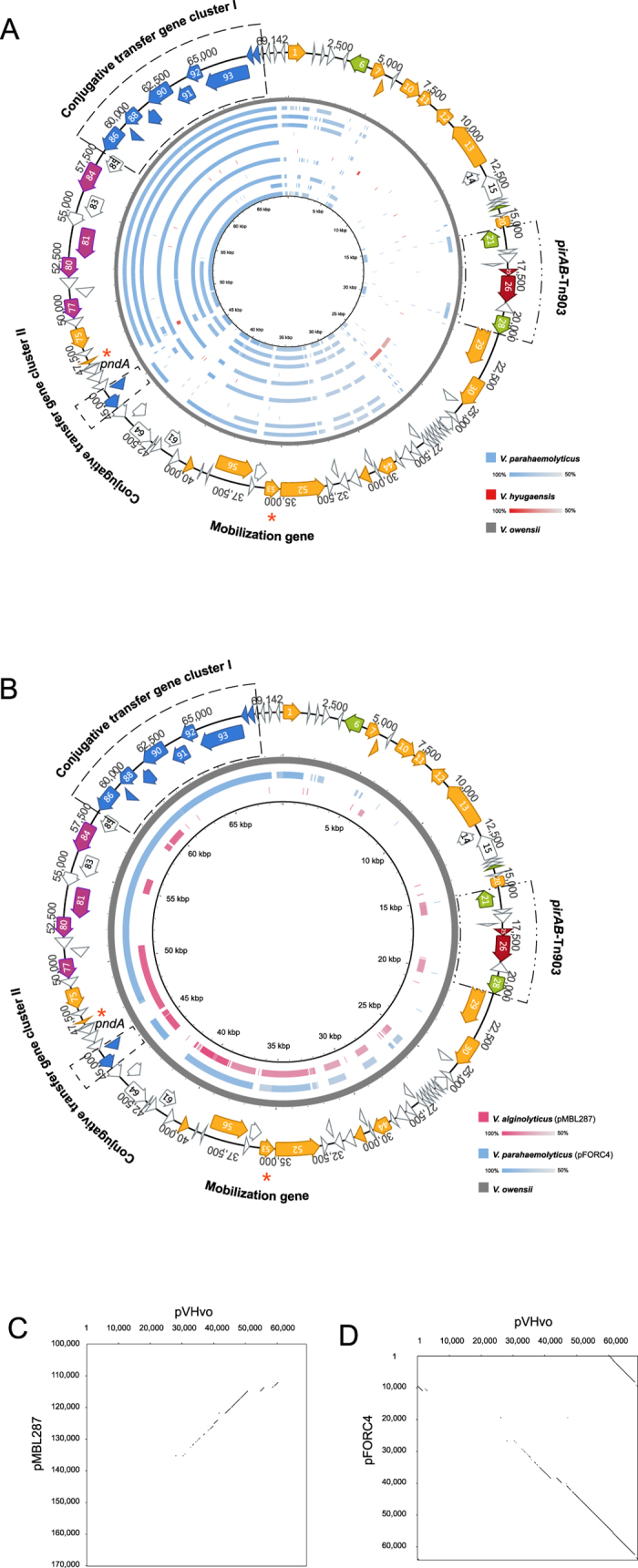
Figure 4. Sequence comparison between pVH and pVH-r.

(A) Sequence identity plot of pVH-r contigs based on comparison to pVHvo. The outer map is the reference sequence map of pVHvo. From outer to inner: pVHvo (gray), AMRZ01000017 (blue), AWHI01000014 (blue), AWIH01000044 (blue), AWJX01000468 (blue), AXNR01000072 (blue), BBLF01000195 (red), JPIP01000006 (blue), JPKV01000063 (blue), JPLU01000004 (blue), JPLV01000074 (blue), and LIRR01000030 (blue). (B) Sequence identity plot of the two complete pVH-r plasmids based on comparison to pVHvo. The outer map is the reference sequence map of pVHvo. The color intensity is proportional to sequence identity. (C) Dot plot comparison of pVHvo and pMBL287 indicating the regions containing shared identical sequence. (D) Dot plot comparison of pVHvo and pFORC4 (partial) indicating the regions containing shared identical sequence.
