
Fig. 1.
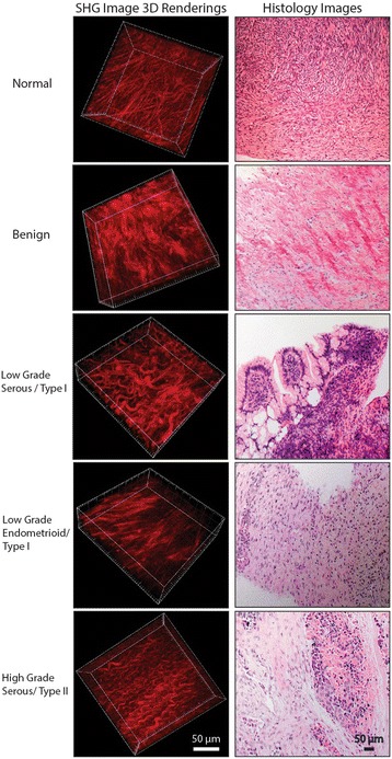
Left column shows 3D renderings of forward directed SHG images of representative normal stroma, benign, LGS, endometrioid, and HGS ovarian tumors obtained at 890 nm excitation. The tissue sections were ~100 μm in thickness. Right column is representative H&E staining of the same tissue. Scale bar = 50 μm