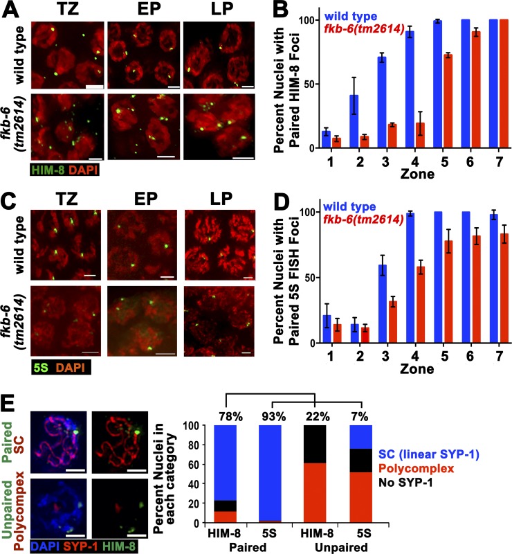
Figure 2.
fkb-6(tm2614) mutants show defects in pairing of the X-chromosome and chromosome V. Representative images of HIM-8 (A) antibody staining or 5S FISH (C) in the genotypes indicated in TZ/zone 3, EP/zone 4, and LP/zone 7. Stained with HIM-8 or FISH (green) and DAPI (red). Images are z-stack projections halfway through the nuclei. Bars, 2 µm. (B) Quantification of percentage of nuclei with paired HIM-8 foci. n nuclei: wild type = 790 and fkb-6 = 614. (D) Quantification of percentage of nuclei with paired 5S foci. n nuclei: wild type = 1,113 and fkb-6 = 1,277. Zones in B and D in terms of meiotic stage are as indicated in Fig. 1 D. Error bars are SD. (E) Representative images of common classes and division into categories of SYP-1 staining with HIM8 or 5S FISH probe (n = 743, 1,028 nuclei). Bars, 2 μm.

