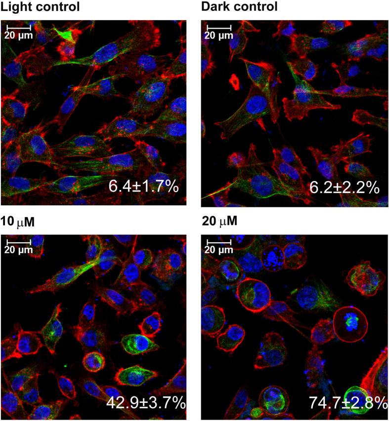
Figure 5. Immunocytochemistry showing cytoskeletal dynamics after PDT with diiodo-squaraine in MDA-MB-231.
Actin stained with Phalloidin TRITC (Red) and Tubulin with anti α-Tubulin antibody and secondary FITC (green) and nucleus Hoechst (blue). Scale bars represent 20 μm. We observed the structural changes in actin cytoskeleton which was spindle shaped in both controls and less ring-shaped actin organisation of about 6.4 ± 1.7% in light control and 6.2 ± 2.2%in dark control. Upon PDT we observed more ring shaped actin about 42.9 ± 3.7% with 10 μM and 74.7 ± 2.8% with 20 μM diiodo-squaraine treatment.

