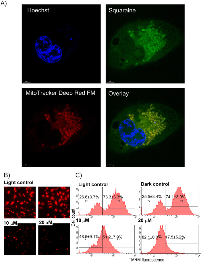
Figure 6. Diiodo-squaraine localizes to mitochondria and induces loss of mitochondrial membrane potential upon PDT.
(A) MDA-MB-231 cells, where treated with 100 μM diiodo-squaraine for 1 hour followed by MitoTracker-far red and Hoechst counter staining shows diiodo-squaraine predominantly localize in mitochondria. Scale bars represent 5 μm (B)The changes in mitochondrial membrane potential were monitored using the Tetramethylrhodamine methyl ester (TMRM), which is a cell-permeant, cationic, red-orange fluorescent dye that is readily sequestered by active mitochondria. The red fluorescence of TMRM is directly proportional to Mitochondrial membrane potential (∆ψM). TMRM shows loss of ∆ψMs after PDT with diiodo-squaraine in MDA-MB-231 cells. a) Fluorescent microscopic images of MDA-MB-231 after PDT with diiodo-squaraine cells showsthe reduced fluorescence of TMRM which is directly proportional to ∆ψM C) Flow cytometric analysis of MDA-MB-231 cells, showing loss of ∆ψM by diiodo-squaraine in PDT. Data are expressed as a mean value ± standard deviation of three independent experiments. Here the population P2 shows background fluorescence, which represents cells with low ∆ψM, and the population P3 shows cells with enhanced fluorescence indicating cells with high ∆ψM. Here we can observe a significant decrease in P3population both at 10 and 20 μM diiodo-squaraine in PDT when compared to both controls.

