Figure 1.
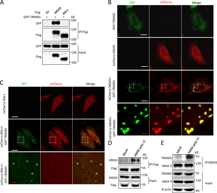
TRIM65 interacts with MDA5. (A) Immunoprecipitation (IP) and immunoblot analysis of the interaction of Flag-MDA5 with GFP-TRIM65 in the lysates of HEK-293T cells. EV, empty vector; input, cell extract without immunoprecipitation. (B and C) Confocal microscopy analysis of HeLa cells cotransfected with plasmids of GFP-TRIM65 with mCherry-MDA5 (B) or mCherry–RIG-I (C). The bottom row shows higher-magnification images of white boxes in the row above. Bars: (white) 20 µm; (red) 2 µm. (D) Immunoblot analysis of the interaction of Flag-TRIM65 with endogenous MDA5 in HEK-293 cells stably expressing Flag-TRIM65 stimulated by HMW poly I:C for 1.5 h, followed by IP with anti-Flag antibody. (E) Immunoblot analysis of the endogenous interaction between TRIM65 and MDA5 in THP-1 cells stimulated by HMW poly I:C for 1.5 h, followed by IP with anti-MDA5 antibody. Data are representative of two (A) or three (B–E) independent experiments.

