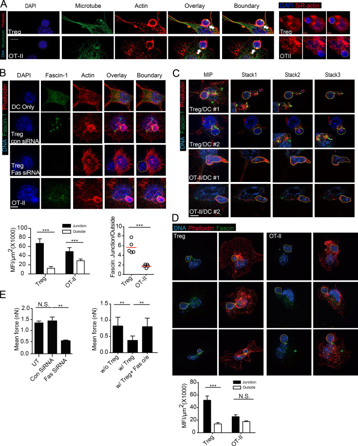
Figure 4.
Polarization of actin and DC-specific actin bundling protein. Fascin-1 is associated with T reg cell–mediated suppression (A) Maximum-intensity projection (MIP) SIM images of a T reg cell or an OT-II T cell coupled with a DC2.4 cell pulsed with OVA, stained for DNA with DAPI, anti-α-tubulin and phalloidin. White arrows highlight the increased thickening of F-actin at the T–DC interface. Hand-traced boundaries of T cells according to DIC are shown to the right here, as well as in the subsequent images. Bars, 5 µm. Representative of at least 10 independent images from four independent stainings. (right) SiR-actin/DAPI-labeled DCs in mixture with T cells (DAPI only) under conventional microscope. (B, top) MIP SIM images of conjugates between T reg cells and DC2.4 cells transfected with control or Fascin-1 siRNA. Conjugates were stained with DAPI, phalloidin, and anti–Fascin-1 antibody. Red boxes covering the T–DC contact sites were used for statistical analyses of Facsin-1 intensity. (bottom) Fascin-1 intensities at T reg cell–DC and OT-II–DC contact sites, as indicated by red boxes in the left. (bottom right) Ratios of mean Fascin-1 intensities in and out of the junction areas. Representative of at least 10 independent images from four independent stainings. (C) MIP SIM images and representative optical sections (stack 1–3) of conjugates between T reg cell (top two rows) or OT-II (bottom two rows) cells and DC2.4 cells. Arrows highlight areas of intermingled Fascin-1 and F-actin bundling, magnified in the inserts. Representative of at least 10 independent images from three independent stainings. (D) As in B, Fascin-1 distribution in three T reg cell–BMDC or OT-II-BMDC pairs was visualized by SIM (top) and quantitatively analyzed in the lower. Representative of three independent stainings. (E, left) Mean forces of OT-II T cells adhering to untreated, control siRNA-treated, or Fascin-1–specific siRNA-treated DC2.4 cells prepulsed with OVA. (right) Mean forces of OT-II T cells adhering to control vector-transfected or Fascin-1–overexpressing DC2.4 cells prepulsed with OVA, with or without T reg cell contact. Each is representative of at least three independent experiments. **, < 0.01; ***, < 0.001; NS, not significant.

