Figure 1.
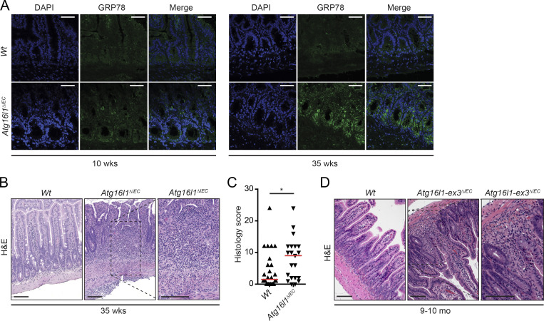
35-wk-old Atg16l1ΔIEC mice exhibit increased ER stress and transmural CD-like ileitis. (A) Representative images of GRP78 (green) immunoreactivity in 10- and 35-wk-old Atg16l1ΔIEC mice. n = 3. DAPI is shown in blue. Bars, 50 µm. (B and C) Representative H&E images (B) and enteritis histology score of 35-wk-old Atg16l1ΔIEC mice maintained in a mouse norovirus–free facility (Cambridge; C). n = 25/21. The median is shown. *, P < 0.05, Mann–Whitney U test. Note the transmural character of inflammation in Atg16l1ΔIEC mice compared with Wt mice. Bars, 100 µm. (D) Representative H&E images of 9–10-mo-old Atg16l1-ex3ΔIEC mice generated and housed at Cedars-Sinai SPF mouse facility in Los Angeles. The inset shows the transmural character of the inflammation seen in Atg16l1-ex3ΔIEC mice. n = 10. Bars, 100 µm.

