Figure 3.
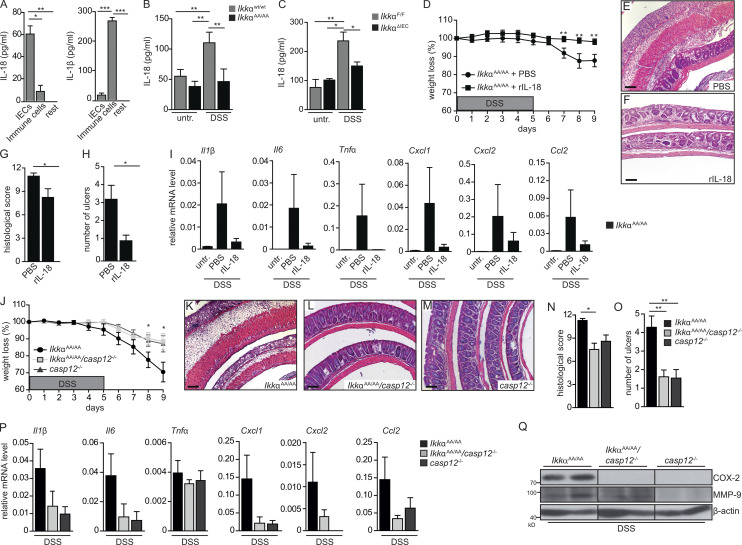
Enhanced caspase 12 activation and decreased cytoprotective IL-18 serum levels in IkkαAA/AA mice. (A) Quantification of ex vivo production of IL-18 and IL-1β by flow cytometry–sorted IECs and immune cells from the colon of WT mice on day 5 of 3.5% DSS treatment. IECs were defined as CD45−, CD11b−, Gr1−, and EpCAM+; immune cells as CD45+, CD11b+, Gr1+, and EpCAM−; and rest as CD45−, CD11b−, Gr1−, and EpCAM−. n = 5. ANOVA followed by Bonferroni posthoc test for multiple datasets was used. (B) Serum IL-18 levels in IkkαWt/Wt and IkkαAA/AA mice on days 0 and 9 of DSS (3.5%) regimen. Data are from two independent experiments. n > 6. ANOVA followed by Bonferroni posthoc test for multiple datasets was used. (C) Serum IL-18 levels were determined by ELISA from IkkαF/F and IkkαΔIEC mice at days 0 and 9 of DSS (3.5%) regimen. n = 5. ANOVA followed by Bonferroni posthoc test for multiple datasets was used. (D) Body weight was determined in IkkαAA/AA mice during DSS (3.5%)-induced acute colitis. Mice were daily i.p. injected either with PBS or recombinant IL-18 (rIL-18; 0.5 µg/d). Data are from two independent experiments. n > 8. Student’s t test was used. (E and F) Representative H&E-stained colon sections from IkkαAA/AA mice treated with PBS (E) or rIL-18 (F) throughout the 3.5% DSS regimen. Bars, 500 µm. (G and H) Histological damage (G) and number of ulcers (H) on day 9 of DSS regimen in IkkαAA/AA mice that had been injected daily either with PBS or rIL-18. Data are from two independent experiments. n > 7. Student’s t test was used. (I) Relative mRNA expression levels of inflammatory mediators in the colon of IkkαAA/AA mice that were left untreated or received either PBS or rIL-18 for nine consecutive days and analyzed on day 9 of the 3.5% DSS regimen. Data are from two independent experiments. n > 3. (J) Body weight was determined in IkkαAA/AA, IkkαAA/AAcasp12−/−, and casp12−/− mice during DSS (2.5%)-induced colitis. Data are from one of two experiments performed. n > 5. Student’s t test was used. (K–M) Representative H&E staining of colon sections from IkkαAA/AA (K), IkkαAA/AAcasp12−/− (L), and casp12−/− (M) mice treated with 2.5% DSS for 5 d and analyzed on day 9. Bars, 500 µm. (N and O) Histological damage (N) and number of ulcers (O) in IkkαAA/AA, IkkαAA/AAcasp12−/−, and casp12−/− mice on day 9 of the DSS regimen. Data are from one of two experiments performed. n > 5. Student’s t test was used. (P) Relative mRNA expression levels of inflammatory mediators in the colon of IkkαAA/AA, IkkαAA/AAcasp12−/−, and casp12−/− mice on day 9 of DSS (2.5%) regimen were determined by qRT-PCR. n > 3. (Q) Whole colonic lysates were prepared from IkkαAA/AA, IkkαAA/AAcasp12−/−, and casp12−/− mice on day 9 of the 2.5% DSS treatment, and protein expression was analyzed by immunoblot analysis using the indicated antibodies. Data are mean ± SEM. *, P < 0.05; **, P < 0.01; ***, P < 0.001. untr., untreated.

