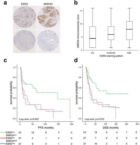
Fig. 4.

SMR3A and ESR2 expression in HNSCC patients. a Representative pictures of immunohistochemical staining (brown signal) of serial tumor sections with anti-ESR2 (left row) or anti-SMR3A antibodies (right row). Haematoxyline counterstaining (blue staining) demonstrates the tissue architecture. Scale bars = 500 μM. b Boxblot depicts the SMR3A immunoreactivity score as mean value and 5th/95th percentile for individual tumors with low, moderate or high ESR2 staining pattern. c-d Kaplan-Meier graphs show differences in disease-specific (DSS) and progression-free survival (PFS) between subgroups without detectable ESR2 staining (ESR2neg, blue line) and ESR2-positive tumors with low (green line) or high SMR3A expression (red line)
