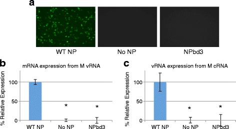
Fig. 3.

NPbd3 is defective for viral protein and RNA expression in reconstituted vRNPs. a Plasmids to express reconstituted vRNPs with GFP-M vRNA and either WT-NP, no NP, or NPbd3 were transfected into 293 T cells. 48 h post transfection cells were observed for GFP-M expression. WT NP represents the positive control while no NP is the negative control. GFP was visualized with a Nikon Eclipse TS100 (Nikon Intensilight C-HGFI for fluorescence) inverted microscope and images captured with the Nikon DS-Qi1Mc camera with NS Elements software. b and c RNA was purified from cells expressing reconstituted vRNPs (b) or cRNPs (c) as indicated. 1 μg was DNase treated and subject to reverse transcription with oligo dT (b) or vRNA specific primers (c) and quantitative PCR with M gene specific primers to calculate relative M RNA expression in each sample. Data are from triplicate trials; asterisks indicate p < 0.02
