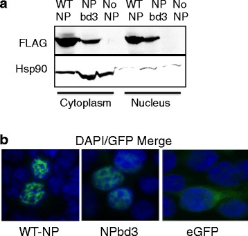
Fig. 4.

NPbd3 is expressed and localized as wild type NP. a Cells expressing reconstituted vRNPs were collected and fractionated with NP-40 non-ionic detergent to break open cellular plasma membrane. Microscopy was used to confirm disrupted plasma membranes and intact nuclei. Nuclei were pelleted by centrifugation and proteins isolated. Proteins were separated on a 10% SDS PAGE gel and transferred to nitrocellulose. Western was performed with anti-FLAG to detect WT NP and NPbd3 and anti-Hsp90 to detect Hsp90, a protein localized in the cytoplasm, which serves as confirmation of cellular fractionation. Expected size was confirmed by comparison with Fisher BioReagents EZ-Run protein standards. b NP-GFP and NPbd3-GFP fusion proteins or eGFP as indicated were expressed in cells grown on poly-L-Lysine cover slips. Cells were washed and fixed using a 1:1 methanol and acetone mixture. The coverslips were mounted onto glass slides using SouthernBiotech™ Dapi-Fluoromount-G™ Clear Mounting Media which stains the cell nucleus blue. Slides were observed on a Nikon ECLIPSE TE2000-U fluorescent microscope and images were captured with an Andor Clara DR-3446 camera using NIS-Elements AR software
