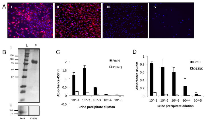
Figure 4. Human uromodulin binding and cell line adherence.
(A) FmlHAD binding to human urothelial cell lines. FmlHAD (i and ii) or K132Q (iii and iv) binding (red) to 5637 (i and iii) or T24 (ii and iv) urothelial cell lines. Hoechst DNA staining is represented by the blue in all panels. (B) FmlHAD far western on urine precipitations. Upper panel (i) represents the SDS-PAGE of precipitated urine where L represents the ladder and P the urine protein concentrate. The lower panel (ii) is a Far Western Blot performed on the samples from (i) with FmlHAD or K132Q mutant. (C–D) ELISA showing FmlHAD and FimH binding to urine precipitate. (C) FmlHAD and K132Q or (D) FimH and Q133K binding to human urine protein precipitates. Columns represent the mean of each data set and bars represent the standard deviation.

