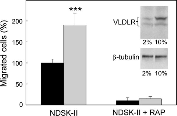
Figure 4. Effect of serum concentration on VLDLR expression levels in HUVECs and NDSK-II-induced migration of neutrophils through HUVEC monolayer.

HUVECs were cultured in the medium containing 2% (black bars) or 10% (grey bars) FBS for 48 hours before transmigration assays. HUVECs were grown to confluency on gelatin-coated cell culture inserts. Calcein AM-labeled HL-60 cells differentiated into neutrophil-like cells were added to the upper chambers on top of the HUVEC monolayers in the presence of 1.5 μM NDSK-II without or with 0.5 μM RAP. The cells migrated into the lower chambers were collected and quantified as described in Materials and methods. The number of cells migrated in the absence of NDSK-II (control) were subtracted and the results were expressed as percentage of the cells migrated in the presence of NDSK-II. The graph shows combined data obtained from 2 independent experiments performed in triplicate; error bars denote means ± SD; ***P < 0.001. Insert shows the levels of VLDLR (top panel) and β-tubulin (lower panel) in HUVECs cultured in 2% or 10% FBS as determined by immunoblotting; upper and lower bands in the top panel represent mature VLDLR and underglycosylated VLDLR precursor, respectively.
