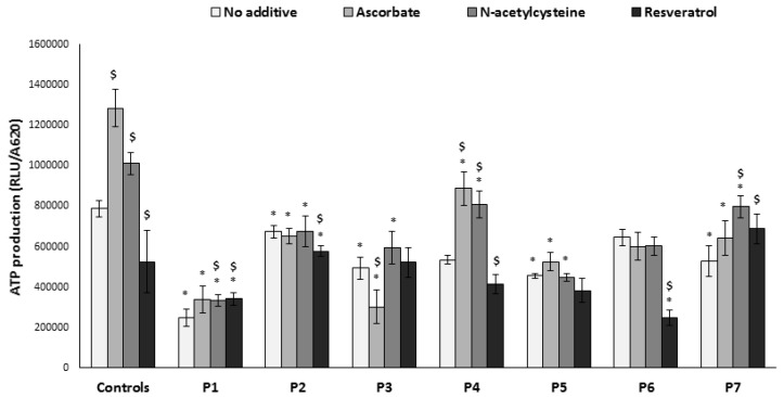
Figure 3.
ATP production. Cells were grown in the absence or presence of the compounds for 72 h in microtiter wells. Subsequently, wells were washed, permeabilized, and incubated with glutamate and malate in the presence of ADP; ATP produced was analyzed by luciferin–luciferase (relative luminescence units, RLU). Results were normalized to cell content measured in parallel wells (A620). Results are presented as mean ± SEM of triplicate wells of at least two independent experiments. * p < 0.05 compared to mean of 3 controls in the corresponding medium, $ p < 0.05 compared to individual patient cells without additive.

