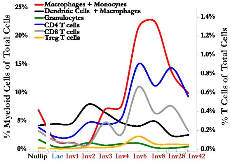
Figure 3.
Immune cells increase in the mammary gland during involution. An influx of immune cells consistent with classic wound healing is observed during mammary gland involution including various myeloid cell populations (left axis): Macrophages and monocytes (CD45+ Gr1intermediate/low F480+ CD11b+, red line); dendritic cells and macrophages (CD45+ CD11c+ MHCII+, black line); and granulocytes (CD45+ Gr1high F480− CD11b+, green line). T cells are also increased in the mammary gland during involution (right axis): CD4 T cells (CD45+ CD3+ CD4+, blue line), CD8 T cells (CD45+ CD3+ CD8+, gray line) and the immunosuppressive Treg T cells (CD45+ CD3+ CD4+ FoxP3+ CD25+, orange line). On the X-axis, the involution window is labeled in red, as “Inv” followed by a number for the day post-weaning. Both axes represent frequencies of indicated cell populations as a fraction of total cells from the gland, as determined by single cell suspensions analyzed by flow cytometry. The Figure is derived from data reported in Reference [45].

