Figure 1.
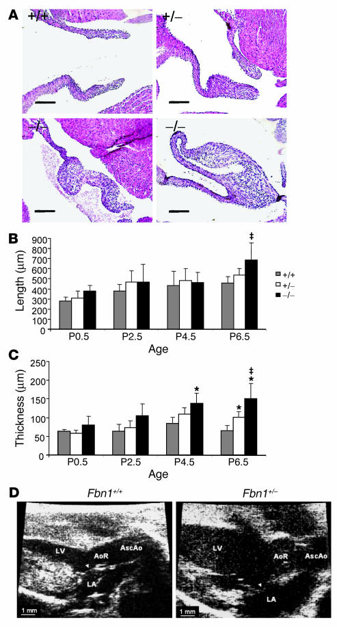
Histologic and morphometric assessment of mitral valve architecture in P6.5 mice. Fbn1 genotypes are indicated as follows: +/+ (Fbn1+/+), +/− (Fbn1C1039G/+), and −/− (Fbn1C1039G/C1039G). (A) Representative mitral valve sections from each genotype at P6.5 showing increased length and thickness in mutant valves as compared to wild-type littermates. Magnification, ×20. Scale bars: 100 μm. (B) Morphometric analysis of mitral valve length during the first week of postnatal life. Fbn1C1039G/C1039G valves were significantly longer by P6.5 when compared with those of Fbn1+/+ animals (–;P –; 0.05). (C) Morphometric analysis of mitral valve thickness during the first week of postnatal life. Increased valve thickness in Fbn1C1039G/C1039G versus Fbn1+/+ mice was significant by P4.5 (*P –; 0.005 vs. Fbn1+/+), and by P6.5, differences between all genotypes were statistically significant (*P –; 0.005 vs. Fbn1+/+; –;P –; 0.05 vs. Fbn1C1039G/+). Error bars indicate 95% confidence intervals. (D) Echocardiographic parasternal long axis views of 9-month Fbn1C1039G/+ and Fbn1+/+ mouse hearts. The anterior mitral valve leaflet (arrowhead) shows increased length and thickening, as well as systolic prolapse into the LA in fibrillin-1–;deficient mice. AoR, aortic root; AscAo, ascending aorta; LA, left atrium.

