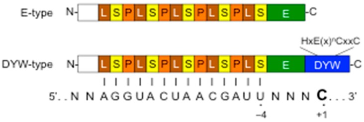
Figure 1.
Plant organellar pentatricopeptide repeat (PPR) editing proteins and a model for their binding to the editing site. Schematic domain structure of PPR editing proteins that consist of PPR motifs (P, L, S), and additional C-terminal domains (E and DYW). The DYW domain contains the conserved zinc-binding motif signature, HxE(x)nCxxC. The PPR tract interacts with a target RNA in a one PPR motif to one nucleotide manner. The last PPR S motif recognizes nucleotide at position –4 from the editing site (+1).

