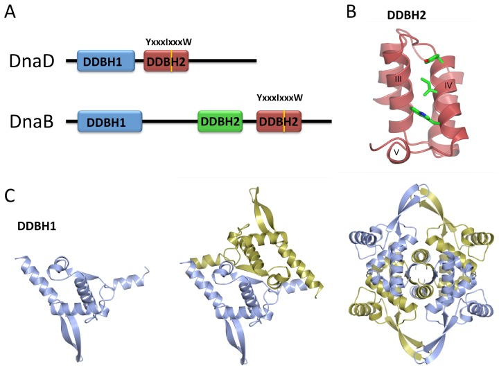
Figure 7.
(A) Diagram showing the architecture of DnaD and DnaB. Conserved DNA binding motif YxxxIxxxW is marked on the relevant DDBH2 domain; (B) Ribbon diagram of the DnaD DDBH2 domain from Streptococcus mutans (PDB code: 2ZC2). Tyrosine, Isoleucine and Tryptophan residues of the YxxxIxxxW motif are coloured by atom (carbon in green, nitrogen in blue and oxygen in red); (C) Ribbon diagram of DnaD DDBH1 domain from Bacillus subtilis (PDB code: 2V79) showing a winged helix with additional structural elements. Monomer, dimer and tetramer architectures are shown. Dimer and tetramer interactions are mediated by the β-strand of the additional helix–strand–helix. Figure inspired by [6].

