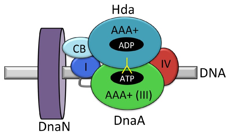
Figure 11.
Schematic representation of the interaction of Hda with DnaA and the β-clamp. Hda-ADP (light and dark cyan) makes contacts with domain I (blue) of DnaA–ATP principally through its clamp binding domain (CB), and with domains III (green) and IV (red) of DnaA through its AAA+ domain. An arginine finger from Hda (yellow) projects into the ATP-binding pocket of DnaA and facilitates ATP hydrolysis as part of the regulatory inactivation of DnaA (RIDA). The DNA-bound β-clamp is shown in purple. Figure adapted from [77].

