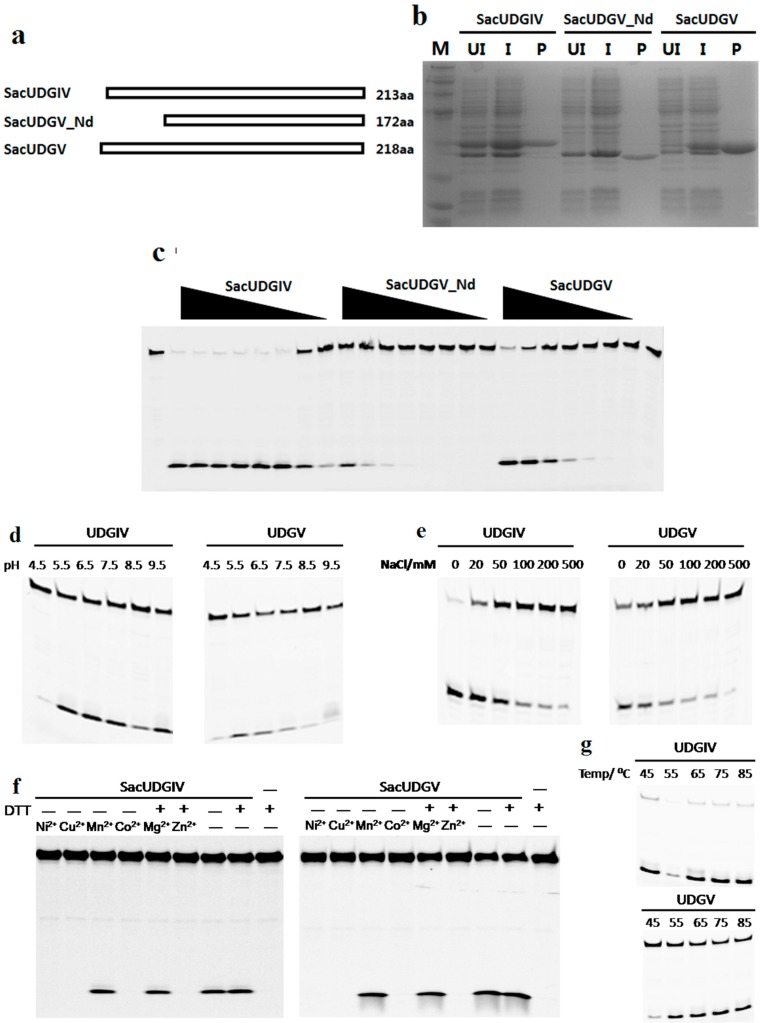
Figure 3.
Uracil removal activities of SacUDGs. (a) Diagram of three UDGs from S. acidocaldarius; (b) 15% SDS-PAGE analysis of recombinant SacUDGs recovered from induced E. coli extracts. The gel was stained with Coomassie blue R-250. Lane M: molecular weight marker; lane UI: uninduced E. coli total proteins; lane I: induced E. coli total proteins; lanes P: purified recombinant protein; (c) Removal of dU from single-stranded DNA by SacUDGs. Approximately 300 ng SacUDGs were diluted in two-fold and incubated with 0.1 μM ss DNA for 15 min at 50 °C. Effect of pH value (d); NaCl concentration (e); divalent ions (f); and reaction temperature (g) on dU removal by SacUDGs. During optimization of reaction conditions about 1 ng SacUDGIV or 10 ng SacUDGV and 0.1 μM dU-carrying single-stranded DNA were incubated at 50 °C for 15 min in assay buffer with various pH value, ion strength, or divalent ions. The reactions were performed at different temperatures for 15 min in an optimal assay buffer.

