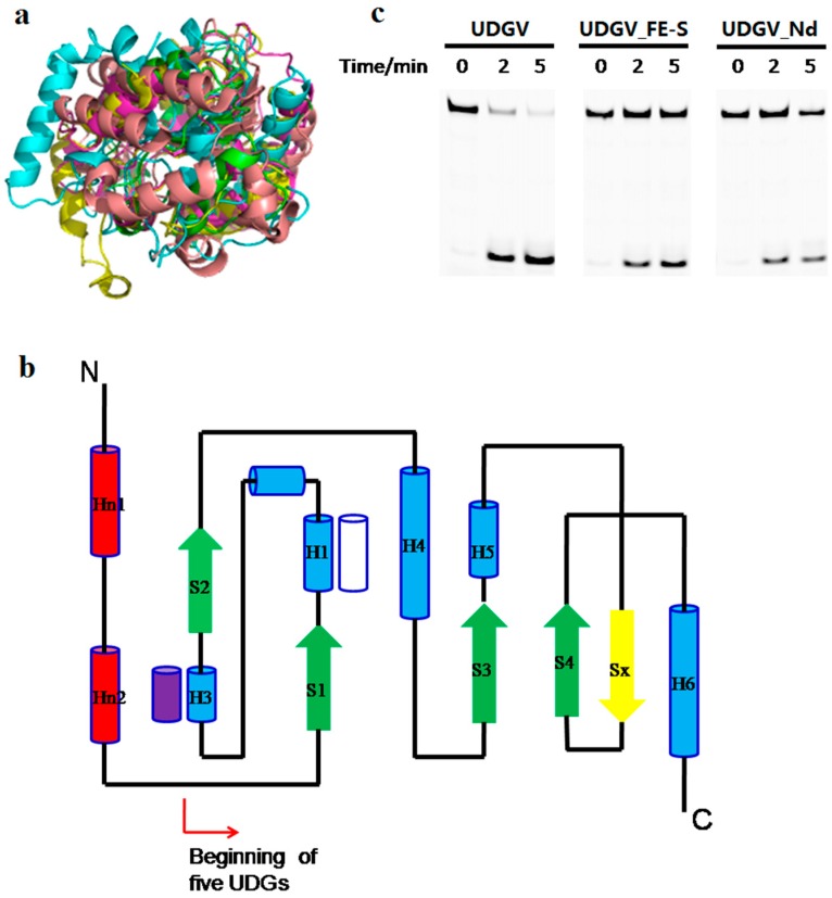
Figure 6.
Structural and sequence multialignment of family 1 to 5 UDGs. (a) Superimposition of five members from UDG superfamily. Each UDG is shown in yellow (TthUDGV, PDB ID: 2d3y), green (EcMUG, PDB ID: 1mug), cyans (EcUNG, PDB ID: 1eug), wheat (GmeSMUG, PDB ID: 5h98), and purple (TthUDGIV, PDB ID: 1ui0), respectively; (b) topology structure of UDG superfamily. The topology of each member of UDG superfamily shows a conserved and unique feature. The secondary-structure elements of the UDG fold are colored as following: common α helices are shown as blue cylinders (the white one is missing in family 1, and the purple one is missing in family 5), and β-strands as green arrows (the yellow one is specific to MUG). The red cylinders are specific to the N-terminal domains of UDGs, excluding the MUG family. (c) Effect of N-terminal helices and [4Fe-4S] on dU removal by SacUDGV.

