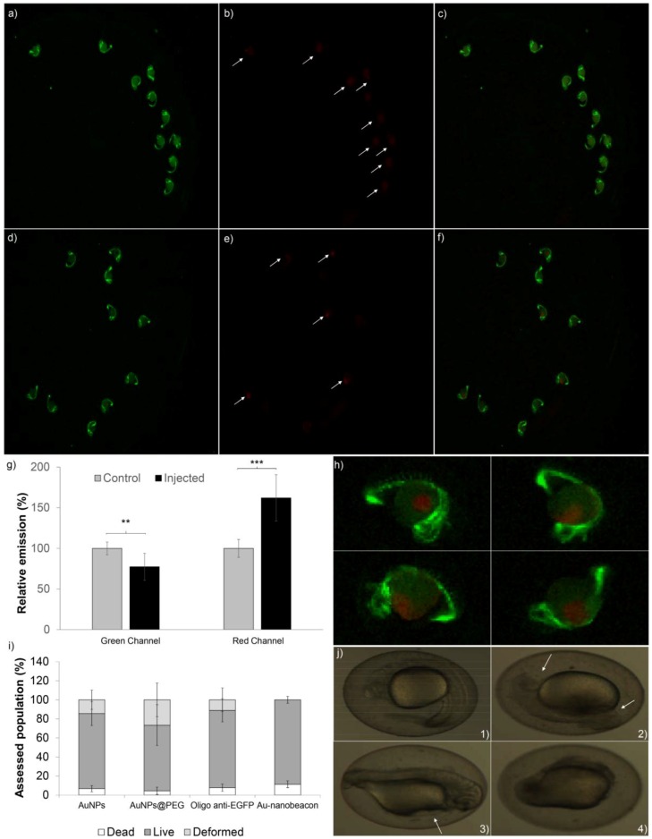
Figure 2.
Au-nanobeacon silencing efficiency of the enhanced green fluorescence protein (EGFP) and acute toxicity assessment. Fluorescence imaging of whole embryos after injection (amplification 8.1×); (a) Green channel of control embryos; (b) Red channel of control embryos; (c) Merged channels for control embryos; (d) Green channel of injected embryos; (e) Red channel of injected embryos; (f) Merged channels for injected embryos (8.1× amplification); (g) Quantification of fluorescence in whole embryos using Image J. The results were normalized to the respective channel of the control. The data are expressed as mean ± standard deviation of five embryos (sample t test—*** for p < 0.05); (h) Zoom of 32.4× of the injected embryos’ merged channels (400% of 8.1×); (i) Quantification of death, survival and morphological malformations upon microinjection of AuNP@citrate, AuNP@PEG, Oligo anti-EGFP and Au-nanobeacon; Error bars corresponds to standard deviation of at least 50 embryos; (j) Example of embryos observed after microinjection of AuNP@citrate, AuNP@PEG, Oligo anti-EGFP and Au-nanobeacon: (j1) Normal embryo; (j2) Head and tail malformation; (j3) Pericardial edema; (j4) Underdeveloped embryo.

