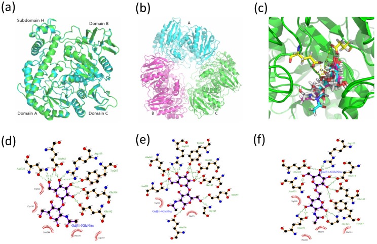
Figure 7.
Three-dimensional structures of MaBGA. (a) Superposition of the MaBGA monomer structure (green) on the structures of β-galactosidase from Thermus thermophilus A4 (cyan; PDB entry 1kwk); (b) Ribbon representation of the trimer structure of MaBGA; (c) Ball and stick representation of the docking models of MaBGA with Galβ1–3GlcNAc (white), Galβ1–4GlcNAc (cyan) and Galβ1–6GlcNAc (magenta), respectively; (d) Schematic diagram of MaBGA/Galβ1–3GlcNAc interactions; (e) Schematic diagram of MaBGA/Galβ1–4GlcNAc interactions; (f) Schematic diagram of MaBGA/Galβ1–6GlcNAc interactions.

