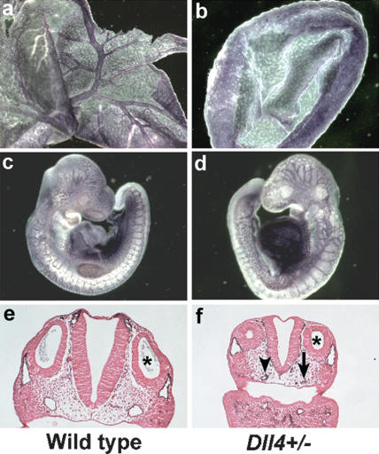
Figure 1.
Vascular defects in Dll4+/- embryos. PECAM-1-stained yolk sacs (a,b) and embryos (c,d) at E9.5. (b) The Dll4+/- mutant yolk sac has failed to remodel the primary vascular plexus to form the large vitelline blood vessels. (d) The vascular network in the Dll4+/- embryo appears less intricate and more primitive than the capillary network of the control littermate. (e,f) Histological sections of PECAM-1-stained E9.5 embryos at the level of the otic vesicle (asterisk). (f) The dorsal aorta of this Dll4+/- embryo is reduced in diameter (arrowhead) on one side and is atretic (i.e., contains no lumen) on the other side (arrow).

