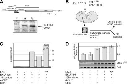
Figure 4.
EKLF is directly involved in the spatial organization of the β-globin locus. (A) Schematic drawing of the EKLF–lbd expression construct used to generate transgenic mice. The Western blot shows expression of the EKLF–lbd fusion protein in the fetal livers of transgenic mice detected by an antibody recognizing the HA tag. (B) Flow chart of the experimental design. Fetal livers are isolated from E12.5 control and EKLF null::EKLF–lbd tg fetuses and disrupted, and the erythroid cells are cultured in the presence of 4-OHT with or without CHX for 16 h. Cells are then harvested, cross-linked with formaldehyde, and subjected to 3C analysis. From a portion of the cells, RNA is isolated to check β-globin gene expression. (C) Expression of β-globin analyzed by real-time RT–PCR. Expression of Hprt was used to standardize the β-globin expression levels. Representative experiment is shown. (D) 3C analysis of the interactions between 5′HS2 and the β-globin promoter. Representative examples of the PCR reactions are shown. Error bars indicate S.E.M. Calreticulin was used as template control.

