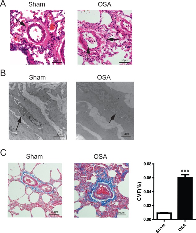
Figure 3. vascular morphological alterations in Sham and OSA canines.
(A) Hematoxylin and eosin (H&E) staining, the arrow indicates the structure change of vascular muscle, scale bar=50μm; (B) Transmission electron microscope images of pulmonary artery smooth muscle, the arrow indicates the ultra-structure change of vascular muscle,scale bar=2μm; (C) Masson's trichrome staining of representative perivascular lung sections and quantification of collagen volume fraction percentage, scale bar=50μm.

