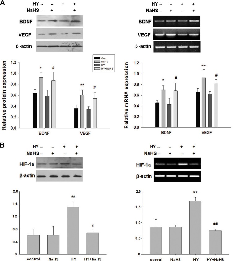
Figure 4. NaHS stimulated the release of BDNF and VEGF, and HIF-1a expression in BMSCs in vitro.
(A) BMSCs exposed to hypoxia-ischemic were incubated in the absence or presence of NaHS (1 μM) for 72 h. The mRNA and protein levels of BDNF, VEGF, and β-actin were then analyzed by western blot and semiquantitative RT-PCR. (B) BMSCs exposed to hypoxia-ischemic were incubated in the absence or presence of NaHS (1 μM) for 72 h. The mRNA and protein levels of HIF-1a and β-actin were then analyzed by western blot and semiquantitative RT-PCR, Each value was normalized to β-actin. Data was expressed as the mean ± SD of n = 4. *p < 0.05, **p < 0.01 NaHS VS Con; #p < 0.05, ##p < 0.01 HY + NaHS VS HY.

