
Figure 1. In vivo treatment of ABC-DLBCL SU-DHL-2 xenografts with OTX015 as a single agent and in combination with other targeted drugs.
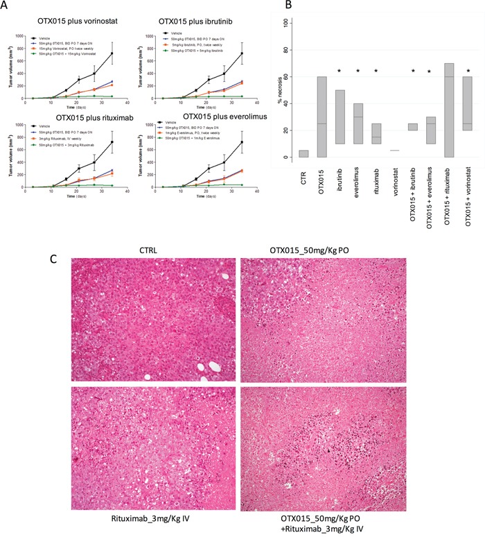
A. Changes in tumor volumes during treatment: Black, vehicle (control mice); Blue; single agent OTX015; Red, single agent targeted drug; Green, OTX015/targeted drug combination. B. Boxplots showing percentage of tumor necrosis at the end of treatment. In each boxplot, the line in the middle of the box represents the median and the box extends from the 25th to the 75th percentile (interquartile range). * P < 0.05 when compared with control (CTR) mice. C. Histopathological analysis revealed control mice or treated only with rituximab displayed vital cell with a diffuse growth pattern (upper and lower left); addition of OTX015 was associated with large areas of coagulative necrosis (Haematoxyln and Eosin, 200X).