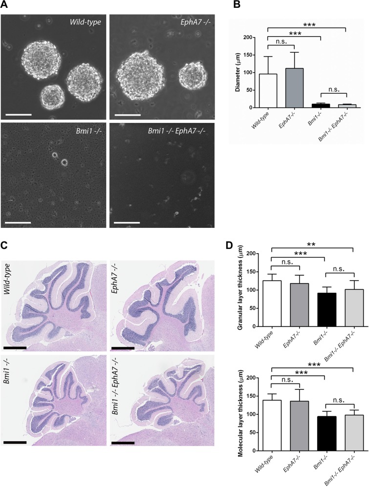
Figure 4. EphA7 deletion does not rescue postnatal neurosphere and cerebellar defects in Bmi1−/− mice.
(A) Neural stem/progenitor cells grown as neurospheres (passage 1). Scale bars 100 μm. (B) Comparison of sphere diameters (n = 10–13). Mean values with standard deviation are shown. In Bmi1−/− and Bmi1−/−/EphA7−/− cultures, most of the cells were single cells and did not form aggregates. (C) Sagittal sections of paraffin-embedded cerebelli stained with hematoxylin-eosin. Scale bars 800 μm. Cells and tissue were isolated from six week-old mice. (D) Granular and molecular layer thickness, mean values with standard deviation. 3 mice per genotype, 3–6 measurements per mouse. Significant results of unpaired t-tests are marked as follows: n.s. not significant (p > 0.05), **p ≤ 0.01, ***p ≤ 0.001.

