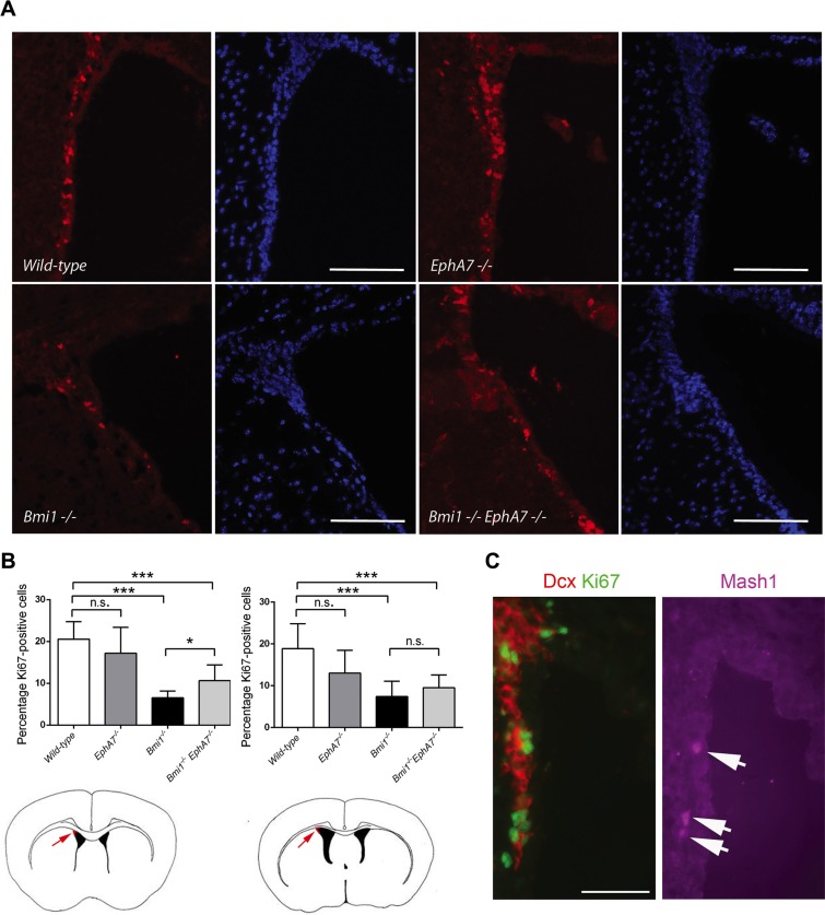
Figure 5. Deletion of EphA7 increases the number of proliferating cells in the LVW dorsolateral corner of Bmi1−/− mice.
(A) Coronal anterior brain sections of 6–7 week-old mice stained with an antibody against Ki67 (red) and DAPI (blue). Scale bars: 100 μm. (B) Number of Ki67-positive nuclei per DAPI-positive nuclei shown as percentages (Wild-type: n = 10 ventricles, EphA7−/− n = 10, Bmi1−/− n = 8, double knockout n = 6). Results of unpaired t-tests are marked as follows: *p ≤ 0.05, ***p ≤ 0.001, ns: not significant (p > 0.05). Mean values with standard deviation are shown. Cartoons depict anterior (left hand side) and posterior (right hand side) forebrain sections. Arrows point to the dorsolateral corner (red). (C) Coronal anterior brain section of a 6 week-old wildtype animal stained with antibodies against Ki67 (green), the neuroblast marker Dcx (red), and the neural progenitor cell marker Mash1 (purple). Arrows point to Mash1-positive cells. Scale bar: 50 μm.

