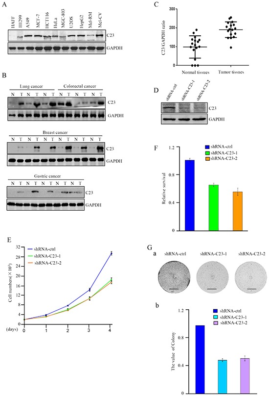
Figure 1. C23 expression levels were elevated in cancer cells and critical for cancer cell proliferation.

A. Protein level of C23 was determined by western blot in different cancer cell lines. GAPDH was used as loading control. B-C. C23 expression levels in human cancer lesions and corresponding normal adjacent tissues were analyzed by Western blot analysis (B) and Graphpad prism (C). D. C23 protein levels were evaluated by western blot in HCT116 cells stably expressing the control shRNA, C23 shRNA1 or C23 shRNA2. GAPDH served as loading control. E. The number of HCT116 cells stably expressing the control shRNA, C23 shRNA1 or C23 shRNA2 was determined by cell counter. The data were represented as mean±S.D. of three independent experiments. F. HCT116 cells stably expressing the control shRNA, C23 shRNA1 or C23 shRNA2 were treated with 100ng/ml Doxorubicin for 24 h. Viability of cells was determined using MTT assays by measuring the absorbance at 490 nm in a microplate reader. G. Long-term colony formation assay of HCT116 cells with and without stable knockdown of C23. Cells (5000 cells per well) were allowed to grow for 3 weeks, then stained and photographed. Scale bar, 1cm.
