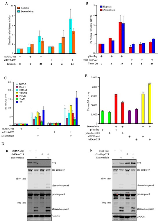
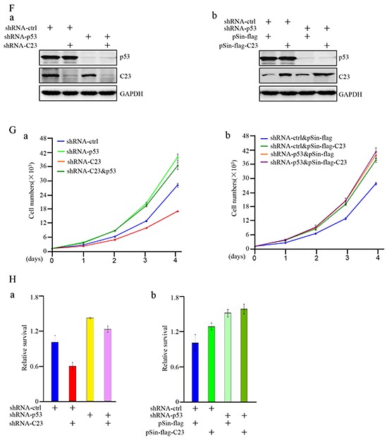
Figure 3. C23 suppressed the transcriptional activity of p53 upon DNA damage and hypoxia.


A. pGL3-3×p53-BS-LUC and Renilla were co-transfected in HCT116 cells combined with and without C23 shRNA. 24 hours after transfection, cells were treated with Doxorubicin (100ng/ml) or upon 1% O2 for indicated times and the cell lysates were analyzed by lucifease assay. B. HCT116 cells with and without stable overexpressing C23 were co-transfected with pGL3-3×p53-BS-LUC and Renilla. 24 hours after transfection, cells were treated with Doxorubicin (100ng/ml) or upon 1% O2 for indicated times and then the cell lysates were subjected to lucifease assays. C. HCT116 cells with and without stable knockdown of C23 were treated with Doxorubicin (100ng/ml) for 24 hours, then p53 target gene mRNA expression levels were analyzed by qRT-PCR analysis. D-E. HCT116 cells with and without stable overexpressing C23 (a) or stable knockdown of C23(b) were treated with Doxorubicin (100ng/ml) or mock control for 24 hours. Cell lysates were then subjected to Western blot analysis with the indicated antibodies (D) and caspase3/7 activity analysis (E), respectively. F. (a) HCT116 cells stably expressing the control shRNA, C23 shRNA, p53 shRNA,or C23 shRNA plus p53 shRNA. (b) HCT116 cells with and without p53 knockdown were introduced with C23 respectively. Protein level of C23 and p53 were evaluated by Western blot. GAPDH served as loading control. G. HCT116 cells stably expressing the control shRNA, C23 shRNA, p53 shRNA,or C23 shRNA plus p53 shRNA (a) and HCT116 cells stably knocked down p53 were additionally introduced with C23 respectively (b). Cell number was evaluated by cell counter. The data were represented as mean±S.D. from three independent experiments. H. (a) HCT116 cells stably expressing the control shRNA, C23 shRNA, p53 shRNA,or C23 shRNA plus p53 shRNA. (b) HCT116 cells stably knocked down p53 were co-transfected with C23 cDNA respectively. Cells were then treated with 100ng/ml Doxorubicin for 24 hours. Cell viability was determined by MTT assay. The data were shown as the mean±s.d. of three independent experiments.
