
Figure 2. ω-3 PUFAs suppress proliferation and induce apoptosis of CRC cells.
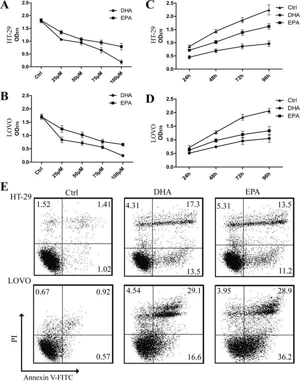
A-D. HT-29 (A) and LOVO (B) cells were assessed by an MTT assay for viability following exposure for 72 h to media containing 10% FBS and varying concentrations of DHA or EPA. HT-29 (C) and LOVO (D) cells were assessed by an MTT assay for viability following exposure for 24h, 48h, 72h and 96h, respectively, to media containing 10% FBS and 75μM DHA or EPA. E. HT-29 and LOVO cells were treated with 75 μM DHA or EPA for 48 h, cell apoptosis was determined by FACS analysis. The data are expressed as the mean ± SEM for triplicate experiments. *P<0.05.