
Figure 6. GPR120, GPR40, Gαs and PKA are involved in mediating ω-3 PUFAs-induced YAP phosphorylation.
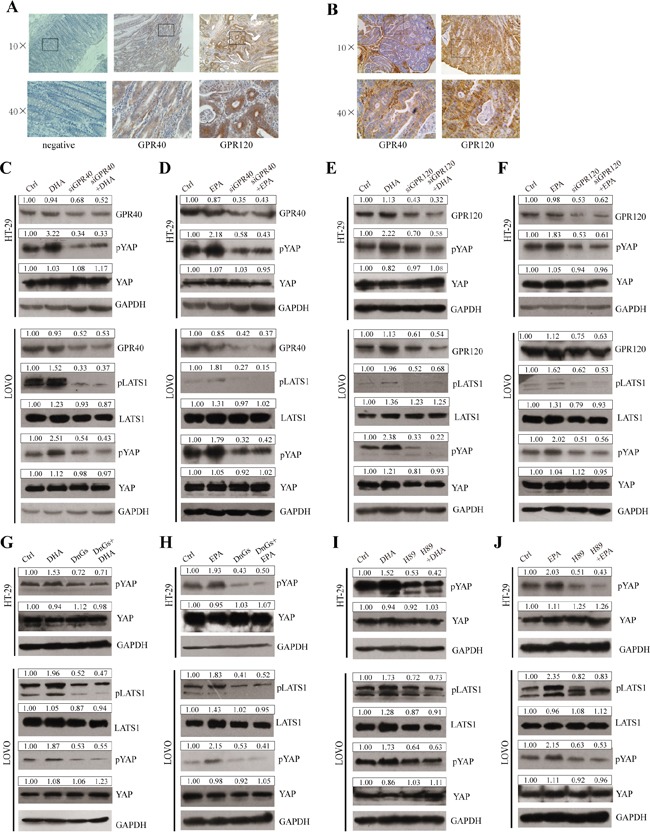
A and B. The expressions of GPR40 and GPR120 in paraffin-embedded CRC tissues of human CRC patients (A) and AOM/DSS-induced mouse modles (B) were examined using IHC staining, 10× and 40×magnification. C and D. GPR40 was knocked down by siRNA and further treated with 75μM DHA (C) or EPA (D) for additional 4h. The expression of pLATS1, LATS1, pYAP, YAP and GPR40 were examined by western blot and quantified in HT-29 and LOVO cells. E and F. GPR120 was knocked down by siRNA and further treated with 75μM DHA (E) or EPA (F) for additional 4h. The expression of pLATS1, LATS1, pYAP, YAP and GPR120 were examined by western blot and quantified in HT-29 and LOVO cells. G and H. Gαs function was blocked by transfected with DnGs and further treated with 75μM DHA (G) or EPA (H) for additional 4h. The expression of pLATS1, LATS1, pYAP and YAP were examined by western blot and quantified in HT-29 and LOVO cells. I and J. PKA was inhibited by the inhibitor H-89 and further treated with 75μM DHA (I) or EPA (J) for additional 4h. The expression of pLATS1, LATS1, pYAP and YAP were examined by western blot and quantified in HT-29 and LOVO cells. GAPDH served as the loading control. Bands were semiquantified by image intensity area under the curve. Intensity of specific band is normalized in relation to loading control protein intensity.