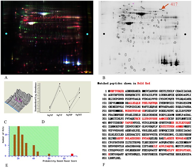
Figure 1. EF2 expression of LSCC tissues and peri-cacinoma lung tissues in 2-D DIGE and MS analysis.

A. Representative image of the overlaid images of Cy- labeled samples. B. Deep-Purple-stained gel image. The number 417 represented the protein spot of interest for EF2. C. The separation effect and protein content for EF2 in samples. The protein content was very high, and the separation effect was good. D. The expressions for protein spot EF2 in non-metastatic and metastatic LSCC tissues respectively, compared with para-carcinoma lung tissues of seven groups. SqNP, SqNT, SqMP and SqMT stand for non-metastastic para-carcinoma lung tissues, non-metastastic lung squamous cell carcinoma, metastastic para-carcinoma lung tissues and metastastic lung squamous cell carcinoma respectively. E. The Mascot score (65) of protein identification for spot 417 by MS. F. A representative tandem mass spectrum of the peptide (in red) matched for EF2 by MS.
