Figure 3.

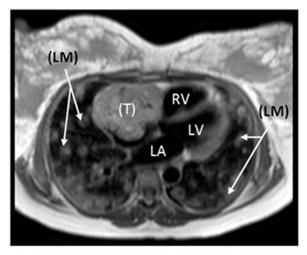
Cardiac MRI revealing large irregular tumor (T) occupying most of the RA, with numerous bilateral lung metastases (LM). RV: right ventricle; LA: left atrium; LV: left ventricle.

Cardiac MRI revealing large irregular tumor (T) occupying most of the RA, with numerous bilateral lung metastases (LM). RV: right ventricle; LA: left atrium; LV: left ventricle.