
Fig 7. Effects of TNFα, IL-1β and LPS on occludin expression and cell viability.
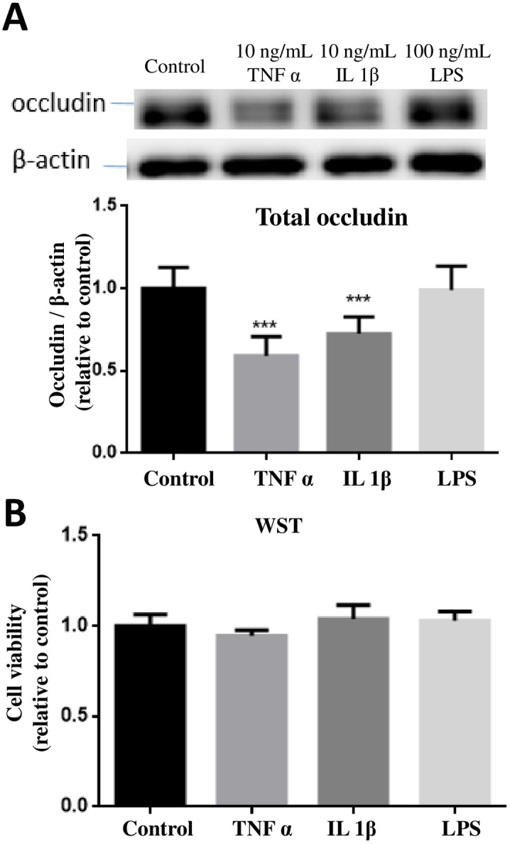
(A) hCMEC/D3 cells were treated with or without TNFα (10 ng/mL), IL-1β (10 ng/mL) and LPS (100 ng/mL) for 24 hours. Cell lysates were collected and occludin and β-actin expression pattern was analyzed by immunoblotting assay. Quantification of the protein band intensity was determined through PItotal/PIβ-actin and then normalized to control. Results are mean ± SD from 4 or more experiments and data are analyzed by one-way ANOVA followed by Bonferroni post-tests (***P<0.001 compared with no treatment control). (B) Cell viability was determined by the WST-1 assay. One-way ANOVA revealed no significant differences among the groups.