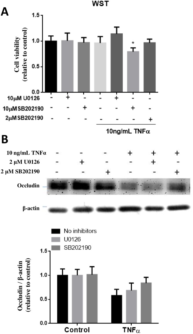
Fig 8. Effects of p-ERK1/2 and p-p38MAPK inhibitors on TNFα-induced occludin expression in hCMEC/D3 cells.
(A) Initial testing for cell viability using WST-1 assay indicated toxicity of cells upon incubation (24h) with TNFα (10 ng/mL) in the presence of SB202190 at 10 μM but not at 2 μM. (B) Testing ability of U0126 (2 μM) and SB202190 (2 μM) to ameliorate the decrease in occludin expression upon exposure of TNFα for 24 h. Cell lysates were collected and occludin and β-actin expression pattern was analyzed by immunoblotting assay. Quantification of protein band intensity was determined through PItotal/PIβ-actin and then normalized to control. Results are mean ± SD from 5 or more experiments. Two-way ANOVA revealed a significant main effect of TNFα (p<0.0001) and the inhibitors (p = 0.0287). Bonferroni post-test showed significant differences between TNFα-treated groups as compared to their respective controls (as indicated by the letter “a”), and TNFα-treated groups with vs. without SB202190 (as indicated by the letter “b”).

