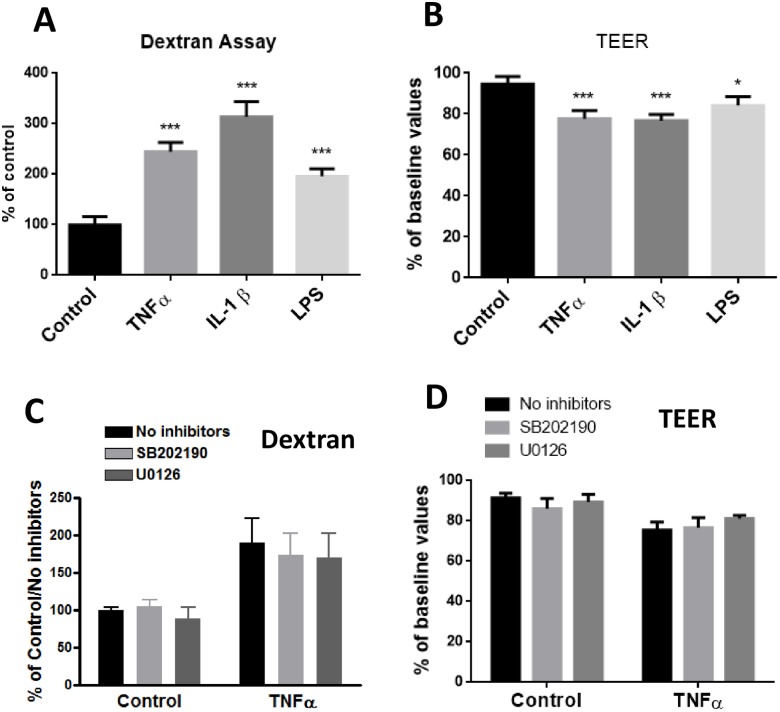
Fig 10. Effects of TNFα, IL-1β and LPS on paracellular permeability as measured by the Dextran and TEER assays.
(A) For the Dextran assay, cells were cultured in inserts for 24 h followed by applying fluorescent FITC-Dextran beads as described in Methods. (B) TEER was determined using an endothelial volt/ohm meter for TEER-EVOM2 as described in Methods. Permeability were determined through FI24h/FI0min (Fluorescence Intensity) and then normalized to control. Results are mean ± SD from 4 or more experiments and data are analyzed by one-way ANOVA followed by Bonferroni post-tests (*P<0.05, ***P<0.001 compared with no treatment control). (C and D) Assessing effects of p-ERK1/2 and p-p38MAPK inhibitors on TNFα-induced changes on paracellular permeability as measured by the Dextran (C) and TEER assays (D). Data are expressed as the mean ± SD of four or more experiments. The results were analyzed by two-way ANOVA, and a significant main effect of TNFα was revealed (p<0.0001 for each). The effects of the inhibitors were not significant.

