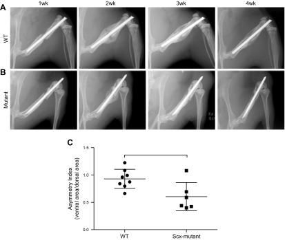
Figure 3.
X-ray images were taken at 7 d intervals after fracture. A) In WT mice, a healing bridged callus was visible at d 14, and the size diminished by d 28, consistent with remodeling. B) In the Scx-mutant mice an asymmetric, partially bridged, smaller callus was evident at d 14 and was less apparent by d 28.

