Figure 1.
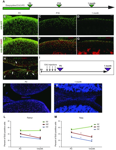
Superficial cells are slow-dividing cells. A) H2B-GFP mice were exposed to doxycycline from E14.5 to P2 and were harvested at P2, P18, and age 1 mo. B–G) The majority of cells were GFP-labeled after exposure to doxycycline (green cells; B, E). The number of labeled cells was reduced markedly after 16 d of chasing (C, F) and only superficial cells still expressed GFP after 1 mo of chasing (D, G). Images in panels E–G correspond to those in panels B–D, but with DAPI (red) channel added. Sample preparation, DAPI staining, laser power, and settings used for confocal scans were similar in B–G. H) To visualize residual GFP, laser power was increased twice compared with that in panel F. Clusters of high (red, arrows) and low (green) proliferative activity could be observed in P18 H2B-GFP mice. I) Between P0 and P2, mice were injected 4 times with EdU, and their bones were collected either on P2 or at age 1 mo. J, K) On P2, many cells throughout the entire articular cartilage were labeled with EdU (J; pink; nuclei were stained with DAPI, blue), whereas only a few cells retained EdU 1 mo later (K). L, M) Quantitative analysis revealed that superficial cells in both the femur (L) and tibia (M) retain EdU, whereas underlying chondrocytes lose this signal. DZ, deep zone; MZ, middle zone; SZ, superficial zone. Values are presented as means ± sem; n = 9. *P < 0.05.

