Figure 2.
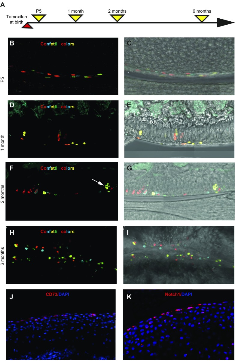
Superficial cells are chondrocyte progenitors. A) PrgCreER(T2):Confetti mice were injected with tamoxifen at birth and analyzed at different timepoints. B–I) At P5, labeled confetti cells (red, yellow, cyan, and, occasionally, green) were observed in the superficial zone of articular cartilage (B, C), whereas labeled chondrocytes were seen clearly 1 (D, E), 2 (F, G), and 6 mo (H, I) after labeling. Large clonal clusters of labeled chondrocytes (arrow in panel F) could occasionally be observed. Images in panels C, E, G, I correspond to those in panels B, D, F, H but with the white light channel added. J, K) Expression of mesenchymal stem cell marker CD73 (J) and Notch1 (K) was observed in superficial chondrocytes. Samples are from 1-mo-old (J) and 5-d-old (K) mice, respectively.

