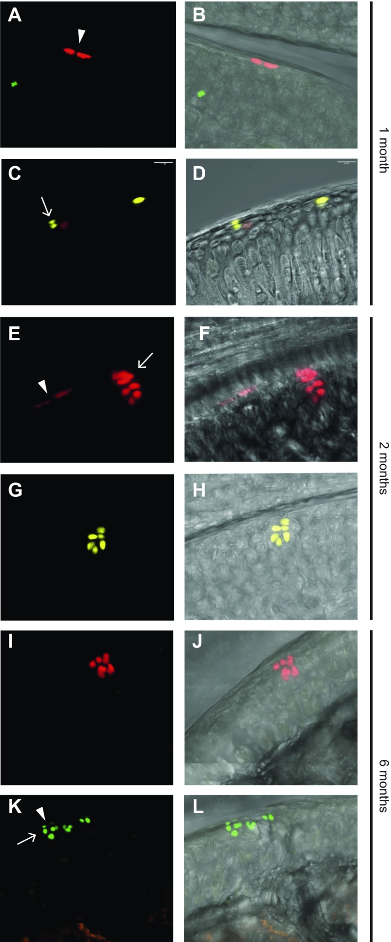
Figure 3.
Cell division in the superficial zone has different orientation. Prg4-traced cells either divided parallel to and then remained at the superficial surface (A, E, K, arrowheads) or divided perpendicularly to this surface (C, E, K, arrows). Clusters of chondrocytes generated from superficial cells can be seen at the cartilage surface together with (E, K) or separated from (G, I) progenitor cells. 3D confocal scans of 150-μm–thick sections were performed to examine the spatial relationship between clusters and the superficial cells above. Images in panels B, D, F, H, J, and L correspond to those in panels A, C, E, G, I, and K but with the white light channel added. Samples for panels A–D were collected at age 1 mo, those for panels E–H were collected at age 2 mo, and those for panels I–L were collected at age 6 mo.

